Research > Focus A > Work Packages A01
Summary
In this project we will further investigate tumor-autonomous communication in glioblastoma cell networks, a central discovery of the first funding period. A small subpopulation of tumor cells located in network hubs exerts periodic, rhythmic Ca2+ oscillations via the potassium channel KCa3.1, continuously activating the glioblastoma network and driving tumor growth. The specific aims are (1) to understand which glioma subtypes depend on this mechanism, (2) how it is influenced by standard therapies, and (3) how KCa3.1 inhibition can be used to break the network-related resistance of glioblastoma towards radio- and chemotherapy and surgical lesioning.
Task 1
Investigating Ca2+ activity in various incurable glioma / glioblastoma types: periodic activity? pacemaking? other patterns?
Steps / Workflow
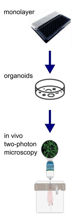
- Establish in vitro monolayer, organoid model, in vivo twophoton microscopy model for other glioma entities (IDH mut., H3K27M; from Unite Core Collection) and more primary glioblastoma models
- Measure and characterize Ca2+ activity across glioma entities (network structure, periodicity, frequencies)
A
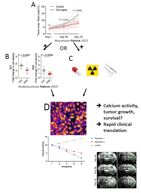
Workflow for testing other glioma entities.
B
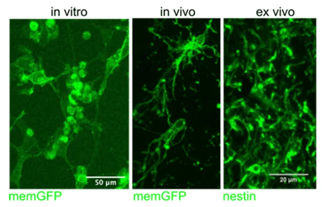
H3K27 mutant cell line in vitro, in vivo, and ex vivo. In vitro and in vivo with membrane GFP fluorescence, immunohistochemistry with nestin staining. Cell lines provided by collaboration with Jones/Witt (A02).
C
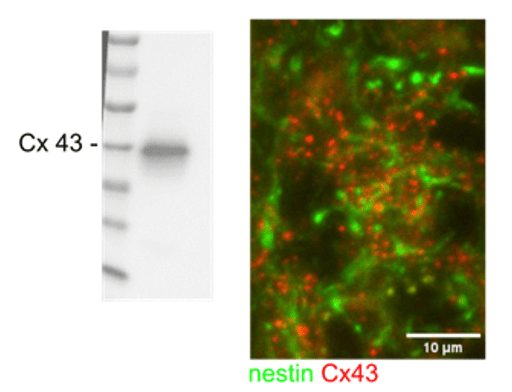
H3K27 mutant cell line expresses high amounts of the gap junction protein Cx43, pointing towards high connectivity. Western Blot from in vitro monolayer, immunohistochemistry stainings of nestin and Cx43 from mouse brain. Cell lines provided by collaboration with Jones/Witt (A02).
D
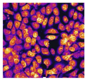
Exemplary image of glioma cells seeded in monolayer for Calcium imaging.
Task 2
Understanding how standard-of-care-treatments (surgical resection, radiotherapy, chemotherapy) alter periodic Ca2+ activity in glioma cells
Steps / Workflow
- Establish pipeline for Ca2+ imaging and analysis after standard-of-care treatments
- Investigate changes in Ca2+ activity after treatments
- Elucidate pathways underlying resistancemediating network communication by scRNA-Seq after using CaProLa methodology (frequency decoding: MAPK, NFkB, others)
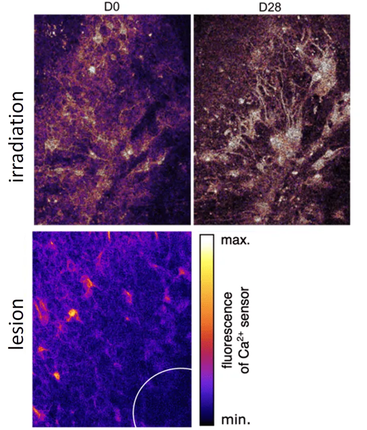
Top: Ca2+ activity of in vivo glioblastoma before (D0) and after (D28) radiotherapy.
Bottom: Ca2+ active glioblastoma cells adjacent to lesion (circle) in vivo. Primary glioblatoma cell line with Twitch3A xenografted into mice, twophoton microscopy image. Circle shows lesion area. Color gradient indicating Ca2+ activity
Task 3
Breaking resistance mediated by network architecture and communication strategy (periodic activity, pacemaking) by KCa3.1 inhibitors
Steps / Workflow
- Perform Senicapoc dose-finding study
- Co-treatment of Senicapoc with cytotoxic therapies to overcome resistance
- Perform co-treatment of Senicapoc and Perampanel for maximum inhibitory effect on both sources of brain tumor Ca2+ activity
- Prepare rapid clinical translation
Previous Funding period

HEUER, SOPHIE, MD
University Hospital Heidelberg, Department of Neurology, Im Neuenheimer Feld 400, 69120 Heidelberg, Germany

WINKLER, FRANK, PROF. DR. MED.
University Hospital Heidelberg, Department of Neurology, Im Neuenheimer Feld 400, 69120 Heidelberg, Germany
Address
Im Neuenheimer Feld 400
69120 Heidelberg
Themen
Research
- Focus A
- A01: Targeting tumor cell network communication to overcome primary and adaptive resistance in glioblastoma
- A02: Development of a specific combination therapy for histone H3-mutant pediatric glioblastoma
- A03: Deciphering resistance against targeted treatments
- A04: Elucidating tumor-associated microglia interactions in astrocytomas CNS WHO-grade 4
- A05: Predictive biomarkers for MGMT-promoter-methylated glioblastoma (2019 – 2023)
- A06: Resistance mechanisms of glioblastoma against alkylating agents and radiotherapy
- A07: Mapping and targeting neuron-tumor networks to tackle therapy resistance in glioblastoma
- A08: Personalized glioblastoma treatment guided by patient-derived tumor organoids
Research
- Focus B
- B01: Mechanisms of response and resistance to glioma-specific t cells
- B02: DNA mis-match repair regulates immune checkpoint blockade therapy in glioblastoma (2019 – 2023)
- B03: Targeting immunosuppressive programs in isocitrate dehydrogenase mutant gliomas
- B04: Impact of myeloid cells on the adaptive immune response in newly diagnosed and recurrent glioblastomas
- B05: Dissecting the response of glioblastoma and its tumor microenvironment to focused high-dose radiotherapy (2019 – 2023)
- B06: Visualization and characterization of immune responses in H3K27M mutant gliomas
Research
- Focus C
- C01: Comprehensive preclinical pharmacology testing of drugs used for glioblastoma treatment
- C02: Radiomics, radiogenomics and deep-learning in neurooncology
- C03: Imaging immune signatures of glioma response and resistance towards immunotherapy (2019 – 2023)
- C04: Metabolic signaling in glioblastoma: a spatial multi-omics approach
- C05: Overcoming glioma radio-resistance with particle therapy
- C06: Functional characterization of EGFR structural variants associated with long-term survival in glioblastoma, IDH-WT


